
Preview
Document Type
Graduate Research
Creation Date
Spring 4-8-2015
Department
Biology
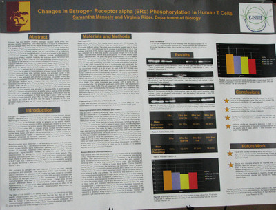
Abstract
Estrogen has two receptor proteins, estrogen receptor alpha (ERa) and estrogen receptor beta (ERJ3). ERa has a half-life of 4 hours for breast cancer and normal target tissue such as the uterus, while ERJ3 has a half-life of 8 hours. The receptor alpha is found to have an importance in breast cancer tumors that are ER-positive. The regulation at the cellular level is key to the effectiveness of endocrine therapies in breast cancer, and an understanding of its underlying mechanism is critical for the identification of novel drug targets for the design of combinatorial therapies. ERa can undergo multiple posttranscriptional modifications (PTMs); however, relatively little is known about the function and regulation of any of the PTMs that ERa can potentially undergo, especially in vivo. Based on the results from two different studies, the structure of ERa for the highest PTMs is the NB domain. In total, 19 phosphorylation sites have been identified in ERa thus far, and most sites contain a Ser-Pro motif. In breast cancer, certain sites of the estrogen receptor (ER) exhibit differential effects of phosphorylation (Ser 104/106, 118, and 167). In some cases phosphorylation of these sites resulted in hypersensitivity to estrogen and an increase in cancerous cell division. In other cases, there was no effect on the progression of breast cancer. Furthermore, different pathways are responsible for th phosphorylation of different sites. These pathways include mitogen-activated protein kinases (MAPK) signaling, IKKa, DK7, a subunit of transcription factor II H, Akt, GSK3J3 p90RSK, mTOR/p70S6K, Rsk, and casein kinase II. Here, I compare phosphorylation of these sites between resting and activated human T cell samples. I purified T cells and extracted the total proteins from both resting T cells and T cells activated with phorbol 12- myrstate 13-acetate (PMA) and ionomycin. I investigated changes in ERa via immunoprecipitation using the ERa antibody and Protein A, as well as Western blot. The blot was reacted with estrogen receptor alpha, phosphor 104/106, 118, and 167 antibodies. These phosphor antibodies only recognize the receptor when the site is phosphorylated. The amount of phosphorylation at each site was compared between resting and activated T cells, and the amount of phosphorylated receptor was adjusted to the total ERa in each sample. Differences between phosphorylation at specific sites in resting versus activated human T cells is expected to reveal potential differences in ERa action and lay the ground work for comparison of these same sites in lupus T cells. The results for a sample size of 10 indicated that when ERa is at 100%, Ser 104/106 resting T cells are 89.30% and active are 92.00%, Ser 118 resting T cells are 80.08% and activated are 87.54%, and Ser 167 resting T cells are 86.44% and activated are 78.35%. These results await statistical analysis.